
Medigest

Endoscopy

NBI: Narrow Band Imaging Endoscopy

Image-enhanced endoscopy that highlights subtle vessel and surface patterns - helping detect early, pre-cancerous changes that are easy to miss.

  • DurationPart of endoscopy / colonoscopy
  • ComfortNo extra discomfort
  • WhereWith endoscopy at the clinic
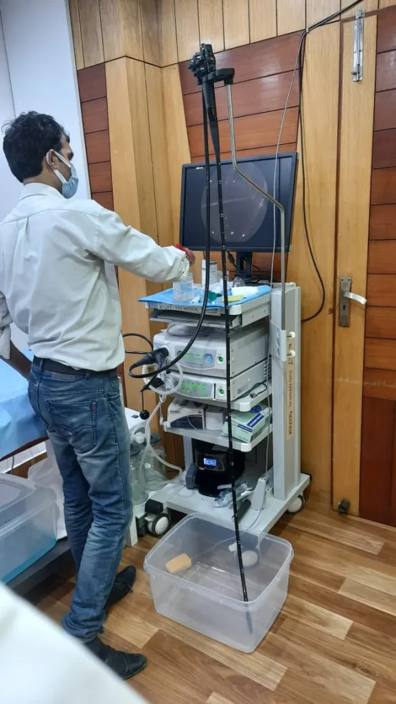
HD video endoscopy system at Medigest clinic

Overview

What is Narrow Band Imaging (NBI) Endoscopy?

Narrow band imaging is an optical filter technology built into modern endoscopes. At the press of a button, it uses specific blue and green light wavelengths that are absorbed by blood, making the blood vessel and mucosal surface patterns stand out clearly.

This helps the doctor characterise lesions in real time, decide where to take biopsies, and detect early cancers and pre-cancerous changes such as Barrett’s oesophagus and flat polyps.

Where: Done at Medigest clinic, West Punjabi Bagh.

Endoscopy

Narrow band imaging endoscopy for early detection

How narrow band imaging works

Normal white light contains all colours. Narrow band imaging filters the light to two narrow bands, blue and green, which are strongly absorbed by haemoglobin. As a result, small blood vessels on the surface appear dark and sharp, and the surface pattern of the lining stands out. Abnormal areas such as early cancers often show irregular vessels and patterns that are easier to recognise with NBI.

Where narrow band imaging is most useful

  • Barrett's oesophagus: to look for areas of dysplasia and target biopsies.
  • Early cancers of the food pipe and stomach: to find and outline subtle lesions.
  • Gastric intestinal metaplasia: a precancerous change that can be mapped.
  • Colon polyps: to predict whether a polyp is likely to be harmless or needs removal.
  • Planning endoscopic treatment: before EMR or ESD.

Narrow band imaging at Medigest

Image-enhanced endoscopy is used during upper GI endoscopy and colonoscopy at Medigest, West Punjabi Bagh, when it helps examine a suspicious area. Dr. Shrihari Anikhindi trained in advanced endoscopy in Japan, where image-enhanced techniques are widely used for early cancer detection.

Does NBI replace biopsies?

No. NBI helps decide where to take biopsies and how to treat a lesion, but the final diagnosis still comes from examining tissue under the microscope.

Why early detection matters

Cancers of the food pipe, stomach and colon are much easier to treat when found early, often with endoscopic removal instead of surgery. Early lesions, however, can be flat and subtle, with only small changes in colour or surface pattern. Image-enhanced endoscopy techniques such as narrow band imaging help the doctor notice these changes and examine them in detail.

Narrow band imaging and polyp assessment

During colonoscopy, NBI helps classify polyps by their vessel and surface patterns. This helps predict which polyps are likely to be precancerous and how they should be removed, and supports planning of EMR or ESD for larger lesions.

Preparing for an NBI examination

No extra preparation is needed beyond that for the endoscopy or colonoscopy. A well-cleaned bowel or an empty stomach is especially important, because good visibility is key to detecting subtle changes.

Book a consultation or request an online consultation to discuss whether Narrow Band Imaging (NBI) Endoscopy is right for you.

Before & after

Who needs it, how to prepare and recovery

General guidance - the clinic gives you personalised instructions when you book.

Why it is done

  • Barrett’s oesophagus surveillance
  • Assessment of polyps in the colon
  • Evaluation of gastritis and intestinal metaplasia
  • Screening in patients at higher risk of GI cancers
  • Targeting biopsies of suspicious areas

How to prepare

  • Same preparation as the endoscopy or colonoscopy being done
  • No additional preparation is needed for NBI

After the procedure

  • Recovery is the same as for the endoscopy or colonoscopy
  • Results are explained along with any biopsy reports

On the day

What happens on the day

  1. 01

    Standard view

    The lining is first examined with high-definition white light.

  2. 02

    NBI view

    NBI is switched on to study suspicious areas in detail.

  3. 03

    Targeted biopsy

    Samples are taken from the most relevant areas.

FAQ

Common questions about Narrow Band Imaging (NBI) Endoscopy

Is NBI a separate procedure?

No. NBI is used during a regular endoscopy or colonoscopy and does not add discomfort or preparation.

Does NBI add time to my endoscopy?

Only a little. NBI is switched on with a button during the procedure, so it adds only a few minutes when the doctor needs to study an area closely.

Who benefits most from NBI?

People with Barrett's oesophagus, a family history of stomach or colon cancer, previous polyps, or suspicious findings on an earlier endoscopy benefit most from a careful image-enhanced examination.

Is NBI safe?

Yes. NBI only changes the colour of the light used by the endoscope. It does not involve dyes, radiation or extra discomfort.

How do I book this test at Medigest?

Book a consultation first by phone, WhatsApp or the online form. Done at Medigest clinic, West Punjabi Bagh. The clinic will share preparation instructions.

Appointments

Consult a gastroenterologist about your digestive or liver symptoms

Visit the clinic in West Punjabi Bagh, or book an online consultation from home.